| Accession: | |
|---|---|
| Functional site class: | PTB ligand |
| Functional site description: | Phosphotyrosine binding (PTB) domains recognize short peptides with a core Asn-X-X-Tyr motif preceded by a short peptide segment that docks by beta augmentation. The classical PTB domains bind the motif when it is phosphorylated on the Tyr residue. However other PTBs recognise essentially the same motif when unmodified. |
| ELMs with same func. site: | LIG_PTB_Apo_2 LIG_PTB_Phospho_1 |
| ELM Description: | This classical version of the motif binds partly as an anti-parallel pseudo beta-strand forming extensive contacts with the beta5 strand and the C-terminal helix of the PTB domain. The peptide itself forms a type 1 beta-turn towards its distal end. This kink is formed by the NPxY motif. The beta-turn positions the tyrosine residue into a localized "anchoring" pocket, where much of the binding energy is concentrated. Additional binding affinity can be achieved by adding one or more hydrophobic residues N-terminal of the core motif at position -5 or nearby. Following the tyrosine residue the peptide turns away from the PTB domain, thereby preventing further contacts. |
| Pattern: | (.[^P].NP.(Y))|(.[ILVMFY].N..(Y)) |
| Pattern Probability: | 0.0001352 |
| Present in taxon: | Metazoa |
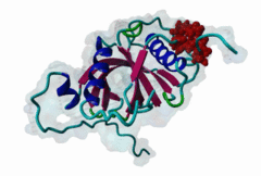
| Interaction Domains: |
|
Phosphotyrosine binding (PTB) domains are structurally conserved modules acting as adaptors or scaffolds to organize signaling complexes involved in a wide range of physiological processes. The PTB domain was first identified in the Shc signaling protein and subsequently in insulin receptor substrate 1 (IRS-1), as a modular domain that recognizes proteins with phosphorylated NPxY motifs. Structural studies have since divided phosphotyrosine-binding PTB domains into Shc-like and IRS-like based on the structure of their peptide binding grooves (Uhlik,2005). More recently, another category of PTB domains was identified as binding to non-phosphorylated (apo) NpxY/F motifs. These phosphotyrosine-independent Dab-like PTBs are thought to represent nearly 75% of proteins encoding PTB domains. The general mode of peptide binding is conserved across all PTB domains. The PTB peptides are bound partly by beta-augmentation of several weakly conserved residues followed by the distal end containing the core consensus motif structured as a beta-turn. There are two fully conserved residues in the NxxY motif but with a strong preference for Proline at the -2 position while there is a weaker preference for a hydrophobic residue at position -5 (positions within PTB motifs are considered relative to the tyrosine residue at position 0.). The combination of both Pro -2 and hydrophobic -5 leads to higher affinity binding. The peptide backbone beta-augmentation further stabilizes the motif interactions. The core phosphorylation-independent motif is exactly the same as the aforementioned motif except with a Phe able to replace the Tyr residue (Nxx[YF]). In contrast to the pTyr motif, the apo-motif is less reliant on the Tyrosine at position 0 and instead relies on hydrophobic interactions and hydrogen bonding along the whole peptide. Furthermore, positions C-terminal of the core motif can contribute to the affinity of the binding. This is in contrast to the phosphorylated motif, that tends to branch away from the PTB surface in the residues following the pTyr. The role of PTB domains is best characterized in the Shc adaptor protein. The presence of both PTB and SH2 domains allows Shc to bind numerous growth factors upon ligand stimulated activation. Upon binding to the activated receptor, Shc itself is phosphorylated and recruits a plethora of adaptor proteins, such as Grb2 and SOS1, leading to downstream pathway activation. To date, Shc is known to bind phosphotyrosine motifs on at least 15 different growth factors or cytokine receptors. Other PTB-containing proteins of the IRS (IRS1-4) and Dok (Dok1-5) families appear to function in a similar way (Uhlik,2005). In contrast to the inducible nature of Shc/Irs/Dok PTB-mediated binding, almost all other PTB domains have specificities independent of phosphotyrosine. In fact, binding of many PTB domains is effectively inhibited by the presence of phosphotyrosine. Several of these Dab-like PTB containing proteins have been linked to Alzeimer's disease on the basis of their binding to amyloid precursor protein (Zhang,1997). These motifs are also strongly associated with endocytic signalling, in particular, receptors containing the motif [FY].NP.[FY] are often endocytosed via adaptor proteins such as Dab1 (Bonifacino,2003). The ability of a motif to bind different PTB domains depending on the phosphorylation state of its tyrosine residue means the motif can act as a phosphotyrosine switch. The NPxY motif in beta-integrin tails alters in this way to regulate the binding of tensin or talin within focal adhesions (Legate,2009). It should also be noted that some, perhaps most, PTB domains have the ability to bind phospholipids. Such a dual role is also found with domains of the PDZ and PH domain families,. The simultaneous binding of phospholipids and peptide to PTB domains has been shown for non-phosphorylated motif binding Dab1-like domains (Howell,1999) whilst phosphopeptides and phospholipids have been reported to mutually compete for binding to the Shc PTB domain (Rameh,1997, Zhou,1995). However, in the later case the recruitment of Shc by the phospholipid precedes phosphopeptide binding (Ravichandran,1997). This ability to bind phospholipids may be crucial for the correct localization of PTB domains to the cytosolic side of cell membrane and in enabling cooperative signalling. Recent studies on the Talin PTB domain have revealed a variant motif with low similarity to the NxxY/F motif, the SPLH motif (Kong,2006). This variant is currently not represented in ELM. |
(click table headers for sorting; Notes column: =Number of Switches, =Number of Interactions)
Please cite:
ELM-the Eukaryotic Linear Motif resource-2024 update.
(PMID:37962385)
ELM data can be downloaded & distributed for non-commercial use according to the ELM Software License Agreement
ELM data can be downloaded & distributed for non-commercial use according to the ELM Software License Agreement