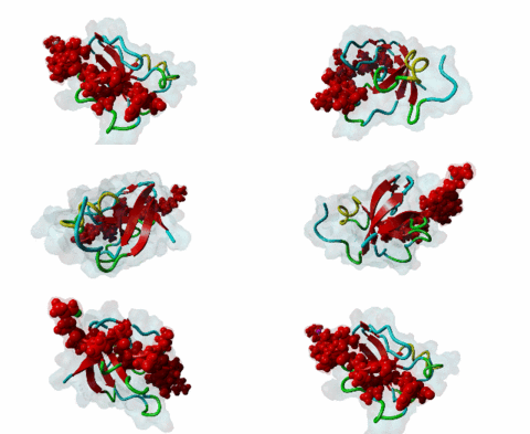
DEG_SCF_FBW7_1
| Accession: | |
|---|---|
| Functional site class: | SCF ubiquitin ligase binding Phosphodegrons |
| Functional site description: | Several phosphodegrons are required for cell state-dependent recognition of regulatory proteins by SCF complexes via repeat domains of associated F box proteins (FBPs) and their subsequent ubiquitin-mediated degradation. The SCF-FBW7 and the SCF-betaTrCP1 motifs, contain two phosphorylated residues, which are recognised via a WD40 domain. For example, the SCF-FBW7 degron TPxxS is found in cyclin E, which is required for the G1/S transition. The SCF-betaTrCP1 degron DSGxxS operates in a broader range of cell regulation. For example, NF-kappa-B inhibitors are phosphorylated and destroyed under immune stimulation while beta-catenin is degraded in the absence of Wnt signalling. Skp2, another FBP, recognises cell cycle regulators via its leucine-rich repeat. In case of the single-phosphorylated DEG_SCF_Skp2-Cks1_1 motif, Skp2 requires additional binding of Cks1 for recognition. So far, only a few cell cycle inhibitors, including p27Kip1 that is mainly involved in G1 arrest, have been found to carry this degron. |
| ELMs with same func. site: | DEG_SCF_FBW7_1 DEG_SCF_FBW7_2 DEG_SCF_SKP2-CKS1_1 DEG_SCF_TRCP1_1 |
| ELM Description: | FBW7 (also called FBXW7, hCdc4 or hSel10) is a member of a family of F-box proteins that binds via WD40 beta propeller to its substrates after their phospho-degron motifs (also named CPDs, i.e. Cdc4 phospho-degrons) have been doubly phosphorylated (Hao,2007, Welcker,2008). The core of the motif is TPxxS, preceded by a variable number of hydrophobic residues. The motif is used in cell cycle regulation: the widely conserved G2 phase-specific cyclin E destruction by FBW7 was first described in yeast. The Thr is often phosphorylated by GSK3, after priming at the other P-site, linking the FBW7 activity with the mitogenic signalling pathway. In some instances the Thr may alternatively be targeted by other kinases such as CDKs. Interestingly, many of the known FBW7 substrates are proto-oncogenes with key roles in the regulation of cell division, differentiation and growth. However, a proposed FBW7 phosphodegron in the key cell state monitor mTor (Mao,2008) does not match the diphosphorylated motif. The phosphodegron in v-Jun is mutated and inactivated, enhancing oncogenicity by preventing its destruction. Some variant motifs substitute a Glu residue for the second phosphosite, e.g. in SV40 large T, and this variant is represented by the alternative pattern in ELM. |
| Pattern: | [LIVMP].{0,2}(T)P..([ST]) |
| Pattern Probability: | 0.0007138 |
| Present in taxon: | Eukaryota |
| Interaction Domain: |
WD40 (PF00400)
WD domain, G-beta repeat
(Stochiometry: 1 : 1)
|
Ubiquitin-mediated proteolysis has diverse regulatory functions in eukaryotic cells (Hershko,1998). The role of ubiquitylation in regulating cell state by targeting proteins for proteasomal destruction is a major field of research. The ubiquitylation of a specific protein is performed by an ubiquitin-protein ligase (also known as E3). Ubiquitin is covalently bound to an ubiquitin-activating enzyme (E1), which transfers the ubiquitin to an ubiquitin-conjugating enzyme (E2). The E2 binds to the E3 ligase, which specifically recognizes the target protein (Kamura,2003). There are two major types of E3 enzymes that ubiquitylate the substrate in different ways: HECT-type E3s first form an E3-ubiquitin thioester conjugate and then transfer the ubiquitin to the substrate. In contrast, RING-type E3s do not form this thioester bond but interact directly with E2 (reviewed in Pickart,2002). The SCF (Skp1-Cullin-F box) is a RING-type E3 consisting of four subunits. These are the scaffold protein Cul1, the RING-domain protein Rbx1/Roc1, the adaptor protein Skp1, and an F box protein that specifically recognises the substrate. F box proteins often contain WD40 beta propeller or leucine-rich repeat (LRR) modules, which interact with one or more short sequences, termed degrons, in their target substrates. Several F box proteins (FBPs), including Cdc4 and Grr1 in yeast, COI1 in plants and betaTrCP, Skp2 and Fbw7 (Fbxw7, hCdc4) in human, recognise phosphorylated degrons in their substrates. Structural and biochemical analysis of three FBPs, betaTrCP, FBW7 and Skp2, improved our understanding of the nature of their interactions with specific substrates. In a first step, single or double phosphorylation of their targets by kinases is required and serves as the ubiquitylation signal. In case of betaTrCP and FBW7 it has been revealed that short phosphodegrons containing two phosphorylated residues bind simultaneously to two phosphate-binding sites on the WD40 domain of the FBPs (Wu,2003; Hao,2007). On the other hand, Skp2 primarily binds substrates via its LRRs. However, in addition to Skp2, the DEG_SCF_Skp2-Cks1_1 motif requires interaction with the accessory Cks1 protein (cyclin-dependent kinases regulatory subunit 1) for its recognition. The Skp2-Cks1 degrons contain only one phosphorylated residue, which binds to Cks1 (Hao,2005). Well characterised phosphodegrons recognised by the ubiquitin ligase system are those of cyclins and cyclin-dependent kinase (Cdk) inhibitors. In yeast, the Cln-cdc28 kinase phosphorylates TPxxS motifs on Sic1 (an inhibitor of the cyclin B regulated kinase) thereby targeting it for degradation and enabling entry into S phase. The FBP Cdc4 recognizes the phosphorylated Sic1. The human ortholog of Cdc4, FBW7, interacts with phosphodegrons of cyclin E, c-Myc and c-Jun (DEG_SCF_FBW7_1; DEG_SCF_FBW7_2). The DEG_SCF-TrCP1_1 destruction motif may play a role in the G2/M checkpoint (e.g. by degrading claspin), but most substrates are regulators of cell state rather than purely responsible for cell cycle. DSGxxS phosphodegrons are found in beta-catenin, several IkB paralogues, Bora as well as the cytosolic side of Prolactin receptor and various other transmembrane receptors. Two members of the Cdk inhibitor protein (Cip/Kip) family, p27Kip1 and p57Kip2, have been shown to contain a functional DEG_SCF_Skp2-Cks1_1 motif, which is recognized by Skp2-Cks1. Skp2 probably recognises other ubiquitylation targets via its LRRs and without supplementary binding of Cks1, e.g. Tob1 and Cdt1 (Zhang,2006). However, recognition of the DEG_SCF_Skp2-Cks1_1 motif requires binding of Cks1 to Skp2. Cks proteins are known as mutation suppressors and regulators of Cdks (Harper,2001) but only Cks1 seems to be able to bind Skp2 and play a role in Cdk inhibitor degradation. p27Kip1 and p57Kip2 (Hao,2005) play a role in maintenance of G1 phase and their degradation occurs during G1/S phase transition. Both proteins specifically inhibit Cdk2-cyclin A or E. In turn, these Cdk-cyclin complexes phosphorylate these Cip/Kip proteins at the motif site as soon as their levels exceed Cdk inhibitor levels in late G1 phase, thereby inducing motif recognition by the Skp2-Cks1 complex and subsequent ubiquitylation and degradation. After mediating relief of CDK inhibition, Cks1 will further regulate CDK activity by acting as a phospho-dependent docking site for cyclin-CDK substrates (DOC_CKS1_1), thereby increasing the specificity and efficiency of substrate phosphorylation by CDK (Koivomagi,2013). The DEG_SCF_Skp2-Cks1_1 motif binds with low affinity to its recognition site since it consists of few amino acids. However, the effective affinity is enhanced by a Cdk-cyclin complex that binds with high affinity across both the substrate targeted for ubiquitylation and the E3 ligase, thereby tightening the motif interaction (Xu,2007). Since Cip/Kip proteins are involved in cell cycle inhibition, they are altered in a broad variety of tumours, e.g. breast and bladder cancers. These alterations mainly comprise reduced expression or increased degradation as well as loss of heterozygosity through locus mutations (Tedesco,2002; Nakayama,1999). Some viruses have been found to use DEG_SCF_TRCP1_1 motifs for perturbation of cellular regulation. A TPxxS-like motif is present in SV40 Large T (ELMI001402) while DSGxxS pseudosubstrate motifs are found in the HIV VPU (ELMI001315) and EBV LMP1 (ELMI001315) proteins and are thought to mop up any available betaTrCP. |
-
Regulation of the cell cycle by SCF-type ubiquitin ligases.
Nakayama KI, Nakayama K
Semin Cell Dev Biol 2005 Jun; 16 (3), 323-33
PMID: 15840441
-
Structure of a Fbw7-Skp1-cyclin E complex: multisite-phosphorylated
substrate recognition by SCF ubiquitin ligases.
Hao B, Oehlmann S, Sowa ME, Harper JW, Pavletich NP
Mol Cell 2007 Apr 13; 26 (1), 131-43
PMID: 17434132
-
Ubiquitin-mediated control of oncogene and tumor suppressor gene products.
Kitagawa K, Kotake Y, Kitagawa M
Cancer Sci 2009 Aug; 100 (8), 1374-81
PMID: 19459846
(click table headers for sorting; Notes column: =Number of Switches, =Number of Interactions)
| Acc., Gene-, Name | Start | End | Subsequence | Logic | #Ev. | Organism | Notes |
|---|---|---|---|---|---|---|---|
| P24864-3 CCNE1 CCNE1_HUMAN |
379 | 384 | SPLPSGLLTPPQSGKKQSSG | TP | 4 | Homo sapiens (Human) | |
| P01106 MYC MYC_HUMAN |
55 | 62 | IWKKFELLPTPPLSPSRRSG | TP | 4 | Homo sapiens (Human) | |
| P05412 JUN JUN_HUMAN |
236 | 243 | QTVPEMPGETPPLSPIDMES | TP | 2 | Homo sapiens (Human) | |
| P24864 CCNE1 CCNE1_HUMAN |
393 | 399 | SPLPSGLLTPPQSGKKQSSG | TP | 3 | Homo sapiens (Human) | |
| P36956 SREBF1 SRBP1_HUMAN |
425 | 430 | KTEVEDTLTPPPSDAGSPFQ | TP | 4 | Homo sapiens (Human) | |
| P13051 UNG UNG_HUMAN |
58 | 64 | PAGQEEPGTPPSSPLSAEQL | TP | 2 | Homo sapiens (Human) |
Please cite:
ELM-the Eukaryotic Linear Motif resource-2024 update.
(PMID:37962385)
ELM data can be downloaded & distributed for non-commercial use according to the ELM Software License Agreement
ELM data can be downloaded & distributed for non-commercial use according to the ELM Software License Agreement

