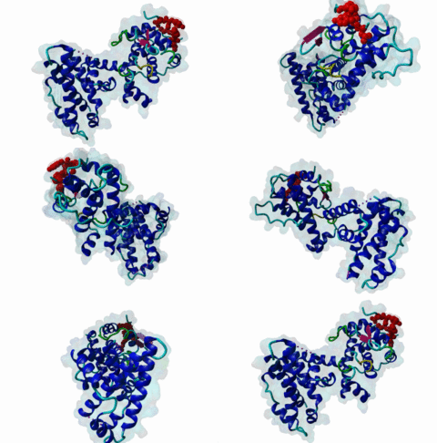
| Accession: | |
|---|---|
| Functional site class: | Pocket protein B Subdomain ligands |
| Functional site description: | Pocket proteins Rb, p107 and p130 play central roles in cell cycle progression and differentiation. The central pocket domain harbors two highly conserved clefts that interact with three different motifs: LIG_RB_pABgroove_1 (LxDLFD), LIG_RB_LxCxE_1 (LxCxE) and LIG_RBL1_LxSxE_2 (LxSxE).The LxDLFD motif binds to a deep groove formed between the A and B subdomains and is present in E2F family transcription factors (E2F1-5). The adenovirus E1A protein mimics the LxDLFD motif and competes with E2F for binding to Rb, promoting E2F activation and cell proliferation. The LxCxE and LxSxE motifs bind to a highly conserved cleft in the B subdomain. The LxCxE motif binds to all pocket proteins and is present in chromatin regulators such as HDAC and KDM5A and in viral proteins. The LxSxE motif is specific for p107/p130 and is present in LIN52, a component of the DREAM complex. A phosphorylation downstream to the core motif acts as a switch that binds to a positively charged pocket only present in p107/p130. |
| ELMs with same func. site: | LIG_RBL1_LxSxE_2 LIG_RB_LxCxE_1 LIG_RB_pABgroove_1 |
| ELM Description: | The LxCxE (LIG_RB_LxCxE_1) motif mediates binding to a highly conserved shallow groove in the B subdomain of pocket proteins (1GUX). The core Leucine, central Cysteine and Glutamic Acid are highly conserved across instances. In a few instances, Leucine is substituted by physicochemically similar Isoleucine. The staggered arrangement, evenly spaced and one residue apart, of the core residues covers one side of an extended, β-strand-like conformation and binds the groove orthogonally, not by β augmentation like many similar staggered motifs (Lee,1998). The Leucine and Cysteine bind into hydrophobic regions of the groove with tight complementarity. The Glutamic Acid forms hydrogen bonds with two backbone amide groups of an α-helix forming one side of the binding groove. The interaction is further stabilised by additional hydrogen bonds to the peptide backbone adding rigidity. Phosphorylation of Rb at Thr821 and Thr826 inhibits LxCxE binding. Several features in the regions flanking the core motif enhance binding affinity. An acidic residue one to three positions preceding the conserved Leucine (optimally D-1) (Jones,1990; Singh,2005) and a hydrophobic residue C-terminal to the conserved Glutamic Acid (optimally L+2) strengthen binding as seen in many viral LxCxE motifs (Singh,2005). Last, an acidic stretch positioned C-terminal to the core motif contributes to fast electrostatically-driven association (Chemes,2011; Chemes,2010). Addition of negative charge by phosphorylation within the acidic stretch further increases binding affinity. The two wild card positions in the core motif also contribute to modulating the binding affinity. In the LxCxE motif from the viral E7 protein, two Tyrosines make stacking interactions that further increase the binding affinity to the low nanomolar range. Cellular LxCxE motifs present a conserved interaction mode and are often suboptimal, with affinities in the micromolar range (Putta,2022). |
| Pattern: | ([DEST]|^).{0,4}[LI].C.E.{1,4}[FLMIVAWPHY].{0,8}([DEST]|$) |
| Pattern Probability: | 0.0005417 |
| Present in taxons: | Metazoa Viridiplantae Viruses |
| Interaction Domains: |
|
Pocket proteins include the paralogs Retinoblastoma (Rb), p107 and p130 in humans (P06400; P28749; Q08999). Pocket proteins play essential roles in cell cycle progression, quiescence and differentiation, and their functional disruption is associated with human cancer. The retinoblastoma susceptibility gene RB1 was the first tumour suppressor gene to be identified and characterised. Inactivation of Rb may contribute to many human malignancies including familial retinoblastoma, small-cell lung carcinomas, cervical carcinomas, prostate carcinomas, breast carcinomas, and some forms of leukaemias (Burkhart,2008). The most studied function of the Rb protein is in the regulation of cell cycle progression at the G1/S boundary (Giacinti,2006). However, Rb is also involved in chromatin remodelling, development, differentiation and apoptosis. Due to the important position of Rb as a regulator of cell cycle progression at the G1/S phase boundary, Rb is highly regulated. Hypophosphorylated Rb binds E2F and recruits histone deacetylases and methyltransferases to repress the expression of E2F controlled gene expression. Phosphorylation by cyclin/CDKs over the course of the G1-phase leads to hyperphosphorylation, disassociation of Rb from E2F and the expression of E2F-controlled S-phase inducing genes (Trimarchi,2002). The Rb paralogs p107 and p130 are closely related and play roles in cell quiescence and differentiation through the formation of the DREAM complex, an evolutionarily conserved transcriptional repressor complex that represses cell cycle genes in quiescent cells and is formed by DP, p107/p130, E2F and the MuvB complex, composed of the core components LIN9, LIN37, LIN52, LIN54 and RBAP48 (Muller,2022). DREAM complex assembly is triggered by LIN52 phosphorylation at Ser28, which allows binding of LIN52 to p107 and recruits hypophosphorylated p107/p130 proteins to MuvB (Fischer,2022). In mammals, MuvB forms the DREAM repressor complex in G0/G1 or the MMB and FOXM1-MuvB activator complexes during S-phase (Muller,2022). E7 disrupts the DREAM complex in HPV-positive cells, leading to increased expression of DREAM target genes, and in vitro disruption of the DREAM complex affects quiescence and induces cell proliferation, increasing the levels of mitotic genes, which is common in high-risk cancers (James,2021; Sadasivam,2013). The multiple roles of pocket proteins are facilitated by its interaction with different protein partners, dependent on the cell type, and on the developmental and cell cycle stages. The interactions of pocket proteins with their binding partners are conserved throughout a wide variety of taxa, from plants to invertebrates and mammals (van den Heuvel,2008). The Rb protein is commonly represented as consisting of three modules, the N-domain, pocket domain and the C-domain (Morris,2001). The pocket domain is further separated into the A and B domains (INTERPRO:IPR002720; INTERPRO:IPR002719) which each possess the helical cyclin fold. The pocket domain acts as a binding region for numerous cellular proteins, including the E2F transcription factors, histone deacetylases and cell cycle regulators as well as viral oncoproteins (Fattaey,1992). The pocket domain structure is conserved in all pocket proteins and harbours two conserved grooves. The first one is a deep groove separating the A and B subdomains that binds to hydrophobic LxDLFD helical motifs (LIG_RB_pABgroove_1) present in the E2F transcription factor transactivation (E2F-TA) domains (1N4M) and the viral E1A protein. The second one is a groove in the B-subdomain that binds to Lx[C/S]xE sequences present in host and viral proteins (LIG_RB_LxCxE_1 and LIG_RBL1_LxSxE_2; 1GUX; 4YOS). These motifs provide functional specificity to pocket proteins through partially understood mechanisms. For example, E2F1/2 show preferential binding to Rb, while E2F4/5 show preferences for p107/p130. While the LxCxE motif binds to all pocket proteins, the LxSxE motif found in LIN52 is specific for p107/p130. The LxCxE motif is found in numerous kinases, histone deacetylases and methyltransferases (e.g. Kim,2001; Lee,2002; Dahiya,2000). Recruitment of histone modifying enzymes to Rb complexes via the LxCxE motif mediates repression of E2F controlled genes. The LxDLFD motif in the transactivation (TA) domain of E2F transcription factors is partially responsible for the recruitment of Rb. Additionally, Rb contacts E2F through a larger disordered region that binds across the E2F/DP1 interface (2AZE). The tight association of Rb to E2F contributes to repressing E2F-mediated transcription. Deregulation of the Rb-E2F interaction or LxCxE-mediated interactions results in hyperproliferation, lack of differentiation, apoptosis, and can lead to cancer. Rb phosphorylation during the G1 phase of the cell cycle produces intramolecular interactions of different Rb regions with the two Rb pocket domain clefts: Phosphorylation of T821 and T826 in Rb induces an interaction of the disordered RbC domain with the pocket domain at a binding site that overlaps substantially with the LxCxE cleft (Rubin,2005). Additionally, phosphorylation of S608 induces an interaction of the flexible linker joining the A and B pocket domains with the E2F binding groove, through mimicry of the LxDLFD motif competing directly with E2F binding (Burke,2012). These intramolecular interactions release E2F transcription factors and induce entry into S-phase. Rb is a common target of viral oncoproteins, predominantly of DNA viruses, most often via the LxCxE motif, which was first identified in the adenovirus E1A and papillomavirus E7 proteins (Jones,1990). Convergently evolved mimics are known in multiple viruses (de Souza,2010) including both plant (RepA in wheat dwarf virus and Clink in Faba Bean Necrotic Yellows Virus) and mammalian (UL97 in Human cytomegalovirus, Large T Antigen in Polyomavirus, E7 in Papillomavirus and E1A in Adenovirus) viral proteins. Viral proteins use their Rb targeting motifs to deregulate E2F binding to Rb, alleviating Rb-mediated repression and forcing the cell into S-Phase thereby activating the replication machinery necessary for completion of the DNA viral life cycle. For example, the LxDLFD motif contained in E1A and E2F use analogous residues to directly compete for the AB pocket of Rb (Liu,2007; 2R7G). Both the canonical E2F and E1A LxDLFD motifs have structural evidence for pocket protein binding (Lee,2002; Liu,2007). Viral proteins may harbour two pocket protein binding motifs. For example, E1A has an LxCxE and an LxDLFD motif which act cooperatively to produce picomolar-affinity binding and E2F displacement (Gonzalez-Foutel,2022). The HPV E7 protein has a candidate LxDLFD motif that binds to Rb (Chemes,2010) but direct biophysical evidence for binding to Rb is still lacking. The short separation between the LxDLFD and LxCxE motifs may prevent simultaneous binding of both motifs from one E7 monomer, but both motifs could contribute to binding of an E7 dimer to Rb. Flanking regions modulate binding affinity and specificity of all pocket protein binding motifs. The structure of the E2F-TA peptide bound to Rb reveals an additional extended interface N-terminal to the LxDLFD motif that creates a high affinity interaction (Lee,2002). The presence of acidic residues N- and C-terminal to the core motif and a fourth hydrophobic position C-terminal to the core motif enhance binding of the LxCxE motif (Palopoli,2018). Phosphorylation of the LxSxE motif acts as a switch that induces binding of LIN52 to p107/p130, recruiting these pocket proteins into the repressive DREAM complex (Guiley,2015). Evidence from binding assays on viral and cellular LxCxE motifs suggests that viral motifs may have evolved for higher pocket protein binding affinities by fine tuning of flanking regions, while host motifs retain suboptimal micromolar affinity binding that may be required for the formation of transient, regulated complexes with pocket proteins (Putta,2022). |
(click table headers for sorting; Notes column: =Number of Switches, =Number of Interactions)
Please cite:
ELM-the Eukaryotic Linear Motif resource-2024 update.
(PMID:37962385)
ELM data can be downloaded & distributed for non-commercial use according to the ELM Software License Agreement
ELM data can be downloaded & distributed for non-commercial use according to the ELM Software License Agreement

