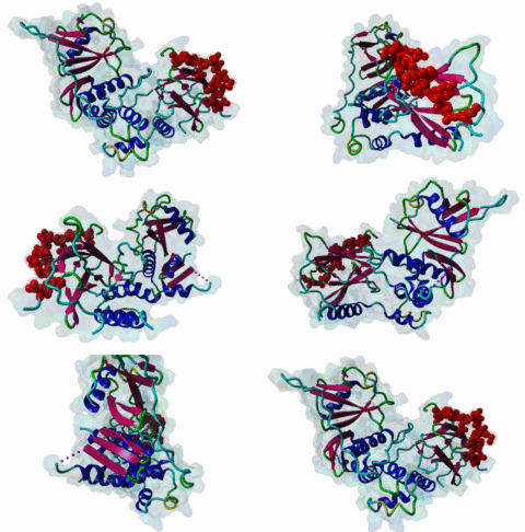
DEG_ODPH_VHL_1
| Accession: | |
|---|---|
| Functional site class: | Hydroxyproline modification in hypoxia signaling |
| Functional site description: | In normoxia, a site in Hypoxia-induced factor (Hif-1) is hydroxylated by prolyl hydroxylases and recognized by von Hippel-Lindau tumor suppressor protein (VHL) which leads to ubiquitination and proteasomal degradation. Under hypoxic conditions, the site is not hydroxylated and thus the protein is not degraded. |
| ELM Description: | The motif of oxygen dependent prolyl hydroxylation is often depicted as LxxLAP. However, that consensus may be misleading as it does not correspond to known interaction preferences. The motif entered in ELM is based on the pVHL interacting residues in the complex with the hydroxylated peptide and their equivalent in vertebrates, fly and worm. It finds a third match in Hif2 alpha in addition to the two experimental motifs |
| Pattern: | [IL]A(P).{6,8}[FLIVM].[FLIVM] |
| Pattern Probability: | 0.0000704 |
| Present in taxons: | Caenorhabditis elegans Drosophila melanogaster Homo sapiens Metazoa Mus musculus |
| Interaction Domain: |
VHL (PF01847)
von Hippel-Lindau disease tumour suppressor protein
(Stochiometry: 1 : 1)
|
Cells respond to decreased oxygen with the induction of an adaptive gene expression programme aimed at restoring oxygen supply and maintaining cell viability during oxygen restriction. Most of the genes induced during low oxygen exposure are under control of the hypoxia inducible factor. The transcription factor Hif-1 (hypoxia-inducible factor 1) plays an essential role in the maintenance of oxygen homeostasis in metazoan organisms. It is a heterodimer composed of an oxygen regulated Hif-1 alpha subunit and a constituvely expressed Hif-1 beta subunit. Under normoxic conditions, Hif-1 alpha is continuously synthesized and degraded, while under hypoxic conditions it accumulates, dimerizes with Hif-1 beta and activates its target gene transcription. In normoxia, Hif-1 alpha is hydroxylated at P402 and P564 (human numbering) by prolyl hydroxylases. This allows binding of von Hippel-Lindau protein (pVHL) which leads to ubiquitination and proteosomal degradation of the Hif-1 alpha . Under hypoxic conditions, hydroxylase activity is reduced due to substrate limitation, inhibition of the catalytic centre or both, this leads to accumulation of Hif-1 alpha which can enter the nucleus, dimerize with Hif-1 beta and activate transcription of hypoxic genes. Activated genes include erythropoietin, glucose transporters, glycolytic enzymes, vascular endothelial growth factor, and others whose protein products increase oxygen delivery or facilitate metabolic adaptation to hypoxia. Hif-1 alpha is often over expressed in the majority of common human cancers and their metastases, due to the presence of intratumoral hypoxia and as a result of mutation in genes encoding oncoproteins and tumor suppressors. Solid tumors require vascularization and so the hypoxia pathway is seen as a major therapeutic target. Three isoforms of Hif alpha (Hif-1 alpha, Hif-2 alpha and Hif-3 alpha) are encoded by distinct genetic loci. Sites from all isoforms align into a conserved LxxLAP motif. This sequence is evolutionarily conserved in Hif alpha proteins from nematodes to mammals (11292861). However, several studies suggest the absence of a rigid consensus sequence for binding of Hypoxia-inducible factor prolyl hydroxylases (Huang,2002; Li,2004). Also, it was shown that downstream residue L574 is required for prolyl hydroxylation and binding of Hif-prolyl hydroxylase 2 (Kageyama,2004). Moreover, the X-ray structure of the complex between Hif-1 alpha peptide bound to pVHL reveals a bipartite binding site, namely, residues 560-567 that include hydroxylated P564 and residues 571-577 that include L574 (1LM8 and 1LQB) (Min,2002; Hon,2002). Two other proteins were also suggested to have the LxxLAP motif. I-kappa-B kinase-beta (IKK beta) protein contains an evolutionarily conserved LxxLAP sequence on a loop of its protein kinase domain. It was suggested that hypoxia releases repression of NF-kappa-B activity through prolyl-hydroxylase dependent hydroxylation of IKK beta. The second protein is the Rpb1 subunit of RNA polymerase II. It was reported that pVHL specifically binds the hyperphosphorylated Rpb1 in a proline-hydroxylation-dependent manner, targeting it for ubiquitination. These finding are difficult to reconcile with the enzymatic studies of prolyl hydroxylase specificity cited above. It is possible that only the Hif1 proteins will prove to have the hypoxia hydroxyproline modification site. |
-
Structure of an HIF-1alpha -pVHL complex: hydroxyproline recognition in
signaling.
Min JH, Yang H, Ivan M, Gertler F, Kaelin WG Jr, Pavletich NP
Science 2002 Jun 7; 296 (5574), 1886-9
PMID: 12004076
-
Structural basis for the recognition of hydroxyproline in HIF-1 alpha by
pVHL.
Hon WC, Wilson MI, Harlos K, Claridge TD, Schofield CJ, Pugh CW, Maxwell PH, Ratcliffe PJ, Stuart DI, Jones EY
Nature 2002 Jun 27; 417 (6892), 975-8
PMID: 12050673
-
Sequence determinants in hypoxia-inducible factor-1alpha for hydroxylation
by the prolyl hydroxylases PHD1, PHD2, and PHD3.
Huang J, Zhao Q, Mooney SM, Lee FS
J Biol Chem 2002 Oct 18; 277 (42), 39792-800
PMID: 12181324
-
HIF prolyl and asparaginyl hydroxylases in the biological response to
intracellular O(2) levels.
Masson N, Ratcliffe PJ
J Cell Sci 2003 Aug 1; 116, 3041-9
PMID: 12829734
-
Leu-574 of human HIF-1alpha is a molecular determinant of prolyl
hydroxylation.
Kageyama Y, Koshiji M, To KK, Tian YM, Ratcliffe PJ, Huang LE
FASEB J 2004 Jun; 18 (9), 1028-30
PMID: 15084514
-
Many amino acid substitutions in a hypoxia-inducible transcription factor
(HIF)-1alpha-like peptide cause only minor changes in its hydroxylation by
the HIF prolyl 4-hydroxylases: substitution of 3,4-dehydroproline or
azetidine-2-carboxylic acid for the proline leads to a high rate of
uncoupled 2-oxoglutarate decarboxylation.
Li D, Hirsila M, Koivunen P, Brenner MC, Xu L, Yang C, Kivirikko KI, Myllyharju J
J Biol Chem 2004 Dec 31; 279 (53), 55051-9
PMID: 15485863
-
Identification of a region on hypoxia-inducible-factor prolyl
4-hydroxylases that determines their specificity for the oxygen
degradation domains.
Villar D, Vara-Vega A, Landazuri MO, Del Peso L
Biochem J 2007 Dec 1; 408 (2), 231-40
PMID: 17725546
(click table headers for sorting; Notes column: =Number of Switches, =Number of Interactions)
| Acc., Gene-, Name | Start | End | Subsequence | Logic | #Ev. | Organism | Notes |
|---|---|---|---|---|---|---|---|
| Q99814 EPAS1 EPAS1_HUMAN |
574 | 585 | IFQPLAPVAPHSPFLLDKFQ | TP | 1 | Homo sapiens (Human) | |
| Q99814 EPAS1 EPAS1_HUMAN |
529 | 542 | LETLAPYIPMDGEDFQLSPI | TP | 1 | Homo sapiens (Human) | |
| Q99814 EPAS1 EPAS1_HUMAN |
403 | 416 | LAQLAPTPGDAIISLDFGNQ | TP | 1 | Homo sapiens (Human) | |
| G5EGD2 hif-1 HIF1_CAEEL |
619 | 631 | LSCLAPFVDTYDMMQMDEGL | TP | 1 | Caenorhabditis elegans | |
| Q9Y2N7 HIF3A HIF3A_HUMAN |
490 | 502 | LEMLAPYISMDDDFQLNASE | TP | 2 | Homo sapiens (Human) | |
| Q24167 sima SIMA_DROME |
1173 | 1184 | NGASIAPVNTKATIRLVESK | TP | 1 | Drosophila melanogaster (Fruit fly) | |
| Q16665 HIF1A HIF1A_HUMAN |
562 | 574 | LEMLAPYIPMDDDFQLRSFD | TP | 3 | Homo sapiens (Human) | |
| Q16665 HIF1A HIF1A_HUMAN |
400 | 413 | LTLLAPAAGDTIISLDFGSN | TP | 1 | Homo sapiens (Human) |
Please cite:
ELM-the Eukaryotic Linear Motif resource-2024 update.
(PMID:37962385)
ELM data can be downloaded & distributed for non-commercial use according to the ELM Software License Agreement
ELM data can be downloaded & distributed for non-commercial use according to the ELM Software License Agreement

