| Accession: | |
|---|---|
| Functional site class: | Helical calmodulin binding motifs |
| Functional site description: | Calmodulin (CaM) is a calcium-dependent regulatory protein known to interact with as many as 320 target proteins. It serves as a primary receptor of intracellular Ca²+ capable of responding to wide range of calcium concentration and translates the Ca²+-signal into a cellular process. CaM is composed of two homologous domains, the N- and C-terminal domains (also called N-lobe and C-lobe), capable of independently folding, connected by a flexible linker. Each domain of CaM contains two helix-loop-helix Ca²+-binding motifs. Upon binding of four Ca²+-ions through these motifs, CaM changes its conformation from a closed form to an open one, exposing a hydrophobic surface capable of interacting with different target proteins. The structural plasticity of CaM allows it to bind different targets with different structural features like protein kinases, phosphatases, receptors, ion-channel proteins, phosphodiesterases, and nitric oxide synthases. The Ca²+-dependent CaM binding site often consists of a basic amphipathic |
| ELMs with same func. site: | LIG_CaM_IQ_9 LIG_CaM_NSCaTE_8 |
| ELM Description: | The IQ motif occurs in myosins and non-myosins proteins and is generally widely distributed in nature. It is sequence with the general consensus [I,L,V]QxxxRGxxx[R,K] with characteristic residues being a hydrophobic residue at position 1, a highly conserved glutamine at position 2, basic charges at positions 6 and 11, and a variable glycine at position 7. Structural studies show that the IQ motif adopts a α-helical conformation, with distinct to no amphipathicity and a net positive charge. The IQ motif enables the Ca2+-independent binding of CaM to target proteins. In some cases (e.g. glycogen phosphorylse b kinase and nitric oxide synthase), the presence of Ca2+ has no effect on CaM-binding, in other cases such as neuromodulin the presence of Ca2+ disables the binding event. The fact that the IQ motif is highly hydrophobic and basic in nature, similar to other Ca2+-CaM binding motifs (LIG_CaM_1-(5-10)-14, LIG_CaM_1-(8)-14 etc.) results in the fact that many IQ motifs can interact with both apo- and Ca2+-CaM. The motif often occurs as multiple tandem repeats in myosins and in some non-myosins proteins. It is sometimes the target of phosphorylation by PKC or PKA proteins. Proteins found to contain at least one IQ domain include myosins, voltage-operated channels, several neuronal growth proteins, phosphatases, sperm surface proteins, Ras exchange proteins, spindle-associated proteins, a RasGAP-like protein and several plant-specific proteins. |
| Pattern: | [ACLIVTM][^P][^P][ILVMFCT]Q[^P][^P][^P][RK][^P]{4,5}[RKQ][^P][^P] |
| Pattern Probability: | 0.0000637 |
| Present in taxons: | Acanthamoeba castellanii Argopecten irradians Bos taurus Branchiostoma lanceolatum Caenorhabditis elegans Capra hircus Carassius auratus Cyprinus carpio Dictyostelium discoideum Drosophila melanogaster Eukaryota Gallus gallus Homo sapiens Macaca fascicularis Macropus eugenii Mesocricetus auratus Monodelphis domestica Mus musculus Oryctolagus cuniculus Papio hamadryas Rattus norvegicus Saccharomyces cerevisiae Schizosaccharomyces pombe Serinus canaria Sus scrofa |
| Interaction Domains: |
|
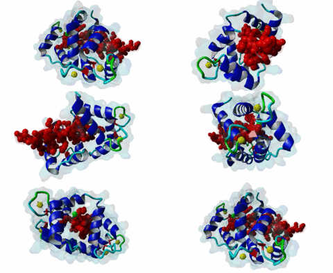
Calmodulin (CaM) is, as its name implies, a calcium modulated protein. It is a small and ubiquitous protein highly conserved in eukaryotes. CaM binds to its target either in a calcium-dependent or -independent manner. The targets of CaM are numerous and diverse as CaM play an important role in many cellular events in animals and plants. Examples of such events are the involvement in ion transportation, several metabolic pathways, cell proliferation, elongation, cell motility, cytoskeleton organization, stress tolerance and transcription (Berchtold,2014). The Ca2+-saturated form of CaM is conformationally different from its apo-form, which leads to the different target specificity of CaM. The apo-form of CaM binds to the IQ motif of its targets, while the Ca²+-bound form binds to a sequence motif, which consists of hydrophobic residues located at specific positions relative to other amino acids. The binding of calcium to the two helix-loop-helix calcium binding motifs in each of the globular domains of CaM induces a conformational change, that exposes a methionine-rich hydrophobic patch on the surface of each domain of CaM, which it used for targeting its binding partners (Gifford,2011). There is no specific consensus sequence for CaM binding in Ca2+-dependent manner. The only common feature is that all binding partners have hydrophobic basic peptides that have the propensity to form an alpha helix. Usually the motif consists of 15-30 amino acids. Based on the positions of the key bulky hydrophobic residues, which make important interactions with the CaM, the motifs can be categorized into different groups, such as 1-14, 1-5-8-14, 1-5-10 and 1-8-14. In the classical mode of binding, the Ca2+-loaded CaM clamps a single helical peptide between the lobes. This is only one of the many possible ways in which CaM interacts with its targets. A growing number of non-classical forms of CaM-binding have been identified, where the conformational changes in the CaM domains differ, so it is able to bind targets in unusual forms, like inverted or in an extended conformation. In other cases binding to the target peptide occurs only via the C-terminal lobe and also the stoichiometry of the complex is not always 1:1, forming more than one alpha helix (Tidow,2013). A Novel group of CaM binding motif, NSCaTE has been identified only in Cav1.2 and Cav1.3 Channels.It is an optional, lower affinity and calcium-sensitive binding site for calmodulin (CaM) which competes for CaM binding with the ancient IQ domain on L-type channels (Taiakina,2013). Studies have shown that the differential binding of CaM is due to its structural flexibility, which arise from the flexible linker-movement between the two CaM domains, and also due to the involvement of a large number of methionine residues found in the exposed hydrophobic patches of CaM (Zhang,1998). Mutations in CaM and also in the CaM-binding regions are associated with many life-threatening conditions. In infants, mutations in CaM affect the calcium signaling events in the heart and lead to cardiac arrhythmias and sudden death of the child (Crotti,2013). Mutations in the CaM-binding region of ryanodine receptors abolish the CaM-binding and induce abnormal hypertrophy with dilatation of the left ventricle, suggesting that CaM-binding is a critical factor for the maintenance of normal function and structure of the left ventricle (Hino,2012). An altered regulation of the CaM-dependent cell cycle and cell proliferation is seen in many tumor cells. Thus targeting CaM or CaM-dependent signalling pathway has been considered a useful strategy for potential therapeutic intervention in cancer. Due to the flexibility of Calmodulin and its variable binding modes, we have not yet been able to derive reliable motifs for the Ca2+ loaded Calmodulin-binding peptides. |
(click table headers for sorting; Notes column: =Number of Switches, =Number of Interactions)
Please cite:
ELM-the Eukaryotic Linear Motif resource-2024 update.
(PMID:37962385)
ELM data can be downloaded & distributed for non-commercial use according to the ELM Software License Agreement
ELM data can be downloaded & distributed for non-commercial use according to the ELM Software License Agreement

