LIG_SPRY_1
| Accession: | |
|---|---|
| Functional site class: | SPRY domain ligand |
| Functional site description: | Peptide motif binding to the members of the SSB (or SPSB) family (SPRY domain- and SOCS box-containing protein). |
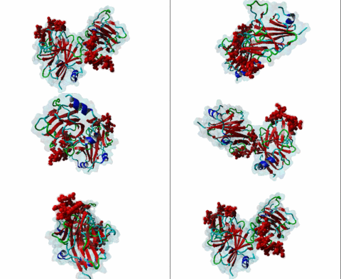
| ELM Description: | Peptides binding to the SPSB -1, -2 and -4 proteins have been identified in hPar-4 (E-L-N-N-NL), for iNOS (D-I-N-N-N), and the Drosophila RNA helicase V-A-S-A (D-I-N-N-N). All three hSPSB proteins share the same conserved SPRY domain fold with two short N- terminal helices packed against a bent β-sandwich, comprising two 7-stranded β-sheets. |
| Pattern: | [ED][LIV]NNN[^P] |
| Pattern Probability: | 7.112e-07 |
| Present in taxon: | Eukaryota |
| Interaction Domain: |
SPRY (PF00622)
SPRY domain
(Stochiometry: 1 : 1)
|
The SPRY domain was first identified in a Dictyostelium discoideum non-receptor tyrosine kinase spore lysis A (splA) and in Mammalia calcium-release channels ryanodine receptors (Ponting,1997). The SPRY domain was subsequently detected in a total of 10 distinct protein families, including the human sequences DDX1, hnRNPs, HERC1 and RanBPM (Ponting,1997). There are currently 2053 eukaryotic proteins listed in the SMART Database (release 6.0) that contain a SPRY domain, including more than 80 proteins encoded in the human genome. Many of these proteins have a conserved N-terminal extension, the PRY domain (50–60 residues) which together with the SPRY domain creates the B30.2 domain (approximately 170 amino acids). It has been shown that the B30.2 domain has evolved recently from the more ancient SPRY domain by incorporating PRY sequences (Rhodes,2005). The B30.2 domain is only found in Vertebrata and it may have been selected as a component of immune defense. The SPRY domain is present in many protein families covering a wide range of functions in different biological processes: the RyR receptors, which regulate intracellular calcium release (Tae,2009); the TRIM5alpha, a host restriction factor acting in the innate immune defense against retroviral infection, including human immunodeficiency virus type-1 (HIV-1) (Zhang,2010); and the SSB family, which is involved in the negative regulation of cytokine signaling (Masters,2005). The SSB-2 is a component of the E3 ubiquitin-protein ligase complex that mediates the proteasomal degradation of target proteins (Kuang,2010). The ~140 amino acid residue domain adopts a novel fold consisting of a β-sandwich structure formed by two four-stranded anti-parallel β-sheets with a unique topology. In combination with the B30.2 domain, these two domains are predicted to adopt a structure resembling the immunoglobulin fold, consisting predominantly of layered anti-parallel β sheets (Seto,1999). |
-
The SPRY domain-containing SOCS box protein SPSB2 targets iNOS for proteasomal degradation.
Kuang Z, Lewis RS, Curtis JM, Zhan Y, Saunders BM, Babon JJ, Kolesnik TB, Low A, Masters SL, Willson TA, Kedzierski L, Yao S, Handman E, Norton RS, Nicholson SE
J Cell Biol 2010 Jul 13; 190 (1), 129-41
PMID: 20603330
(click table headers for sorting; Notes column: =Number of Switches, =Number of Interactions)
| Acc., Gene-, Name | Start | End | Subsequence | Logic | #Ev. | Organism | Notes |
|---|---|---|---|---|---|---|---|
| Q96IZ0 PAWR PAWR_HUMAN |
68 | 73 | PAAAAANELNNNLPGGAPAA | TP | 1 | Homo sapiens (Human) | |
| P09052 vas VASA1_DROME |
184 | 189 | RRRRNEDDINNNNNIVEDVE | TP | 1 | Drosophila melanogaster (Fruit fly) |
Please cite:
ELM-the Eukaryotic Linear Motif resource-2024 update.
(PMID:37962385)
ELM data can be downloaded & distributed for non-commercial use according to the ELM Software License Agreement
ELM data can be downloaded & distributed for non-commercial use according to the ELM Software License Agreement

