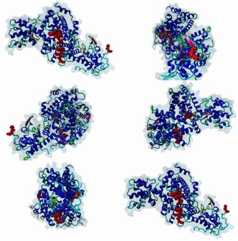
| Accession: | |
|---|---|
| Functional site class: | Pocket protein B Subdomain ligands |
| Functional site description: | Pocket proteins Rb, p107 and p130 play central roles in cell cycle progression and differentiation. The central pocket domain harbors two highly conserved clefts that interact with three different motifs: LIG_RB_pABgroove_1 (LxDLFD), LIG_RB_LxCxE_1 (LxCxE) and LIG_RBL1_LxSxE_2 (LxSxE).The LxDLFD motif binds to a deep groove formed between the A and B subdomains and is present in E2F family transcription factors (E2F1-5). The adenovirus E1A protein mimics the LxDLFD motif and competes with E2F for binding to Rb, promoting E2F activation and cell proliferation. The LxCxE and LxSxE motifs bind to a highly conserved cleft in the B subdomain. The LxCxE motif binds to all pocket proteins and is present in chromatin regulators such as HDAC and KDM5A and in viral proteins. The LxSxE motif is specific for p107/p130 and is present in LIN52, a component of the DREAM complex. A phosphorylation downstream to the core motif acts as a switch that binds to a positively charged pocket only present in p107/p130. |
| ELMs with same func. site: | LIG_RBL1_LxSxE_2 LIG_RB_LxCxE_1 LIG_RB_pABgroove_1 |
| ELM Description: | The LxDLFD motif (LIG_RB_pABgroove_1) mediates binding to a highly conserved deep groove formed in the interface between the A and B subdomains of the Rb, p107 and p130 pocket domains. The motif adopts a helical conformation with three hydrophobic positions facing the base of the groove (Liu,2007; Lee,2002). The first hydrophobic position allows [LIMVA], the second allows [LMF] and the last position favours aromatic residues, with preference for [FY]. The conserved acidic residue between the first and second hydrophobic residues makes extensive charge contacts with the pocket domain, with D424/425 in E2F1/2 and E45 in E1A forming a salt bridge with R467 in Rb. The conserved last acidic position D48 from E1A (2R7G) makes extensive hydrogen bonds with the main chain of S644/T645 and the hydroxyl moiety of S644/S646. The corresponding position in E2F1/2 (D428/429) faces the solvent and does not form interactions, suggesting one additional residue in helical conformation, as observed in E1A, is required for proper positioning of the last acidic residue. In E1A this last residue (L49) makes main chain hydrogen bonds with H44, stabilising the helix. Thus, we include a final wild-card position, although the role of this position on binding affinity has not been tested. Upon phosphorylation of S608 in a flexible linker of Rb, this “RbLoop” mimics the LxDLFD groove motif, competing with E2F binding. A structure of the phosphomimetic S608E mutant (4ELL) reveals a conserved helical structure, conserved positioning of hydrophobic residues and conserved interactions for the two acidic positions (D604 and E608) (Burke,2012). The phosphorylated RbLoop binds intramolecularly to the Rb pocket domain (Burke,2010). An additional hydrophobic [IL] position is present and makes contacts to the Rb pocket. Extensive interactions in the N-terminal flanking region of E2F increase Rb-binding affinity (Lee,2002). It is not yet known if an equivalent (but divergent) motif exists outside Metazoa. |
| Pattern: | ..[LIMVA].[DE][LMF][FYM][IL]{0,1}([DE]|(S)). |
| Pattern Probability: | 0.0000508 |
| Present in taxons: | Metazoa Viruses |
| Interaction Domains: |
|
Pocket proteins include the paralogs Retinoblastoma (Rb), p107 and p130 in humans (P06400; P28749; Q08999). Pocket proteins play essential roles in cell cycle progression, quiescence and differentiation, and their functional disruption is associated with human cancer. The retinoblastoma susceptibility gene RB1 was the first tumour suppressor gene to be identified and characterised. Inactivation of Rb may contribute to many human malignancies including familial retinoblastoma, small-cell lung carcinomas, cervical carcinomas, prostate carcinomas, breast carcinomas, and some forms of leukaemias (Burkhart,2008). The most studied function of the Rb protein is in the regulation of cell cycle progression at the G1/S boundary (Giacinti,2006). However, Rb is also involved in chromatin remodelling, development, differentiation and apoptosis. Due to the important position of Rb as a regulator of cell cycle progression at the G1/S phase boundary, Rb is highly regulated. Hypophosphorylated Rb binds E2F and recruits histone deacetylases and methyltransferases to repress the expression of E2F controlled gene expression. Phosphorylation by cyclin/CDKs over the course of the G1-phase leads to hyperphosphorylation, disassociation of Rb from E2F and the expression of E2F-controlled S-phase inducing genes (Trimarchi,2002). The Rb paralogs p107 and p130 are closely related and play roles in cell quiescence and differentiation through the formation of the DREAM complex, an evolutionarily conserved transcriptional repressor complex that represses cell cycle genes in quiescent cells and is formed by DP, p107/p130, E2F and the MuvB complex, composed of the core components LIN9, LIN37, LIN52, LIN54 and RBAP48 (Muller,2022). DREAM complex assembly is triggered by LIN52 phosphorylation at Ser28, which allows binding of LIN52 to p107 and recruits hypophosphorylated p107/p130 proteins to MuvB (Fischer,2022). In mammals, MuvB forms the DREAM repressor complex in G0/G1 or the MMB and FOXM1-MuvB activator complexes during S-phase (Muller,2022). E7 disrupts the DREAM complex in HPV-positive cells, leading to increased expression of DREAM target genes, and in vitro disruption of the DREAM complex affects quiescence and induces cell proliferation, increasing the levels of mitotic genes, which is common in high-risk cancers (James,2021; Sadasivam,2013). The multiple roles of pocket proteins are facilitated by its interaction with different protein partners, dependent on the cell type, and on the developmental and cell cycle stages. The interactions of pocket proteins with their binding partners are conserved throughout a wide variety of taxa, from plants to invertebrates and mammals (van den Heuvel,2008). The Rb protein is commonly represented as consisting of three modules, the N-domain, pocket domain and the C-domain (Morris,2001). The pocket domain is further separated into the A and B domains (INTERPRO:IPR002720; INTERPRO:IPR002719) which each possess the helical cyclin fold. The pocket domain acts as a binding region for numerous cellular proteins, including the E2F transcription factors, histone deacetylases and cell cycle regulators as well as viral oncoproteins (Fattaey,1992). The pocket domain structure is conserved in all pocket proteins and harbours two conserved grooves. The first one is a deep groove separating the A and B subdomains that binds to hydrophobic LxDLFD helical motifs (LIG_RB_pABgroove_1) present in the E2F transcription factor transactivation (E2F-TA) domains (1N4M) and the viral E1A protein. The second one is a groove in the B-subdomain that binds to Lx[C/S]xE sequences present in host and viral proteins (LIG_RB_LxCxE_1 and LIG_RBL1_LxSxE_2; 1GUX; 4YOS). These motifs provide functional specificity to pocket proteins through partially understood mechanisms. For example, E2F1/2 show preferential binding to Rb, while E2F4/5 show preferences for p107/p130. While the LxCxE motif binds to all pocket proteins, the LxSxE motif found in LIN52 is specific for p107/p130. The LxCxE motif is found in numerous kinases, histone deacetylases and methyltransferases (e.g. Kim,2001; Lee,2002; Dahiya,2000). Recruitment of histone modifying enzymes to Rb complexes via the LxCxE motif mediates repression of E2F controlled genes. The LxDLFD motif in the transactivation (TA) domain of E2F transcription factors is partially responsible for the recruitment of Rb. Additionally, Rb contacts E2F through a larger disordered region that binds across the E2F/DP1 interface (2AZE). The tight association of Rb to E2F contributes to repressing E2F-mediated transcription. Deregulation of the Rb-E2F interaction or LxCxE-mediated interactions results in hyperproliferation, lack of differentiation, apoptosis, and can lead to cancer. Rb phosphorylation during the G1 phase of the cell cycle produces intramolecular interactions of different Rb regions with the two Rb pocket domain clefts: Phosphorylation of T821 and T826 in Rb induces an interaction of the disordered RbC domain with the pocket domain at a binding site that overlaps substantially with the LxCxE cleft (Rubin,2005). Additionally, phosphorylation of S608 induces an interaction of the flexible linker joining the A and B pocket domains with the E2F binding groove, through mimicry of the LxDLFD motif competing directly with E2F binding (Burke,2012). These intramolecular interactions release E2F transcription factors and induce entry into S-phase. Rb is a common target of viral oncoproteins, predominantly of DNA viruses, most often via the LxCxE motif, which was first identified in the adenovirus E1A and papillomavirus E7 proteins (Jones,1990). Convergently evolved mimics are known in multiple viruses (de Souza,2010) including both plant (RepA in wheat dwarf virus and Clink in Faba Bean Necrotic Yellows Virus) and mammalian (UL97 in Human cytomegalovirus, Large T Antigen in Polyomavirus, E7 in Papillomavirus and E1A in Adenovirus) viral proteins. Viral proteins use their Rb targeting motifs to deregulate E2F binding to Rb, alleviating Rb-mediated repression and forcing the cell into S-Phase thereby activating the replication machinery necessary for completion of the DNA viral life cycle. For example, the LxDLFD motif contained in E1A and E2F use analogous residues to directly compete for the AB pocket of Rb (Liu,2007; 2R7G). Both the canonical E2F and E1A LxDLFD motifs have structural evidence for pocket protein binding (Lee,2002; Liu,2007). Viral proteins may harbour two pocket protein binding motifs. For example, E1A has an LxCxE and an LxDLFD motif which act cooperatively to produce picomolar-affinity binding and E2F displacement (Gonzalez-Foutel,2022). The HPV E7 protein has a candidate LxDLFD motif that binds to Rb (Chemes,2010) but direct biophysical evidence for binding to Rb is still lacking. The short separation between the LxDLFD and LxCxE motifs may prevent simultaneous binding of both motifs from one E7 monomer, but both motifs could contribute to binding of an E7 dimer to Rb. Flanking regions modulate binding affinity and specificity of all pocket protein binding motifs. The structure of the E2F-TA peptide bound to Rb reveals an additional extended interface N-terminal to the LxDLFD motif that creates a high affinity interaction (Lee,2002). The presence of acidic residues N- and C-terminal to the core motif and a fourth hydrophobic position C-terminal to the core motif enhance binding of the LxCxE motif (Palopoli,2018). Phosphorylation of the LxSxE motif acts as a switch that induces binding of LIN52 to p107/p130, recruiting these pocket proteins into the repressive DREAM complex (Guiley,2015). Evidence from binding assays on viral and cellular LxCxE motifs suggests that viral motifs may have evolved for higher pocket protein binding affinities by fine tuning of flanking regions, while host motifs retain suboptimal micromolar affinity binding that may be required for the formation of transient, regulated complexes with pocket proteins (Putta,2022). |
(click table headers for sorting; Notes column: =Number of Switches, =Number of Interactions)
| Acc., Gene-, Name | Start | End | Subsequence | Logic | #Ev. | Organism | Notes |
|---|---|---|---|---|---|---|---|
| P06400 RB1 RB_HUMAN |
600 | 609 | PLQNNHTAADMYLSPVRSPK | TP | 8 | Homo sapiens (Human) | |
| P03255 Early E1A 32 E1A_ADE05 |
41 | 49 | SHFEPPTLHELYDLDVTAPE | TP | 6 | Human adenovirus 5 | |
| Q15329 E2F5 E2F5_HUMAN |
333 | 341 | FNLDDNEGVCDLFDVQILNY | TP | 1 | Homo sapiens (Human) | |
| Q16254 E2F4 E2F4_HUMAN |
400 | 408 | YNLDESEGVCDLFDVPVLNL | TP | 2 | Homo sapiens (Human) | |
| O00716 E2F3 E2F3_HUMAN |
442 | 450 | SLGEEEGISDLFDAYDLEKL | TP | 1 | Homo sapiens (Human) | |
| Q14209 E2F2 E2F2_HUMAN |
420 | 428 | GLEAGEGISDLFDSYDLGDL | TP | 6 | Homo sapiens (Human) | |
| Q01094 E2F1 E2F1_HUMAN |
419 | 427 | GLEEGEGIRDLFDCDFGDLT | TP | 9 | Homo sapiens (Human) |
Please cite:
ELM-the Eukaryotic Linear Motif resource-2024 update.
(PMID:37962385)
ELM data can be downloaded & distributed for non-commercial use according to the ELM Software License Agreement
ELM data can be downloaded & distributed for non-commercial use according to the ELM Software License Agreement

