| Accession: | |
|---|---|
| Functional site class: | GLEBS Motif |
| Functional site description: | The GLEBS binding motif is an important cell cycle regulatory motif involved in the spindle checkpoint. |
| ELM Description: | The Gle2-binding-sequence (GLEBS) motif binds to the WD40 domains of Gle2, Bub3 and BubR1 and possibly other paralogous proteins. The first Glu/Asn is important for the overall backbone geometry and makes some water contacts. The second position is a hydrophobic core packing residue. The third residue caps the first helix. The pair of adjacent glutamic acids are deep in the pocket of the binding domain and are the key binding residues. The following residue is important for the core helix. |
| Pattern: | [EN][FYLW][NSQ].EE[ILMVF][^P][LIVMFA] |
| Pattern Probability: | 9.069e-07 |
| Present in taxons: | Eukaryota Homo sapiens Mus musculus Saccharomyces cerevisiae |
| Interaction Domain: |
WD40 (PF00400)
WD domain, G-beta repeat
(Stochiometry: 1 : 1)
|
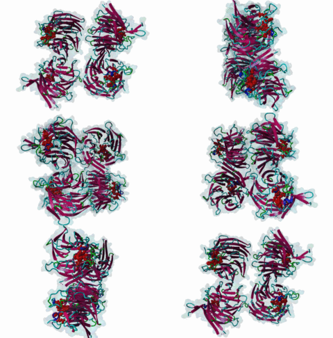
The cell cycle is the process through which a cell replicates itself resulting in two identical daughter cells. The progression of the cell through this process is monitored by a number of checkpoints (Wells,2004). These cell cycle checkpoints halt, abort or delay the progression of the cell through the cycle until certain events, requirements or processes have correctly taken place. Cell cycle arrest protein Bub3 is an important cell cycle checkpoint protein controlling the response of the cell to incorrect spindle formation. Mitotic checkpoint protein Bub3, together with mitotic arrest deficient like protein Mad2, mitotic spindle checkpoint protein Mad3, and cell division cycle homolog protein Cdc20 forms part of the spindle assembly checkpoint, which acts as an inhibitory complex at the kinetochore, preventing the progression of mitosis (Hardwick,2000). The spindle assembly checkpoint is important for regulating the correct segregation of chromosomes to daughter cells during mitosis, by monitoring the progression from metaphase to anaphase. Mad3 in yeast is an integral, but not essential, part of the spindle checkpoint. When it detects defects in the kinetochore and microtububle interactions in the mitotic spindle it arrests the transition, allowing the defective sister chromatid pairs to correctly link up. This ensures the correct segregation later. Proper segregation of the chromatids is achieved when the kinetochores of the two sister chromatids are attached to opposite spindle poles. The acetylation of Bub1 has been shown to regulate its degradation and therefore the inactivation of the spindle assembly checkpoint. In yeast, Bub3 is a substrate for the protein kinase Bub1 (Roberts,1994), and interacts physically with Mad3 (Hardwick,2000). Homologs of the yeast Bub3 in Drosophila, mouse and human have been identified and most have been shown to localise to unattached kinetochores. Cdc20 interacts with Mad2 in the anaphase-promoting complex to inhibit the transition to anaphase (Rudner,1997). This pathway involves the Cdc20-mediated ubquitination of securin. Securin blocks Separase function, delaying the degradation of the cohesin complex, which is a large complex that forms around the sister chromatids after replication. The degradation of this complex allows the separation of the sister chromatids. BUBR1 is involved in the inhibition of the anaphase-promoting complex/cyclosome (APC/C) by blocking the binding of CDC20 to APC/C, independently of its kinase activity. Shin,2004 state that BUBR1 may also be involved in triggering apoptosis in polyploid cells that exit aberrantly from mitotic arrest. The GLEBS motif was identified in yeast nuclear porin Nup116 docking with nuclear pore protein Gle2 (Bailer,1998). The GLEBS motif found in mammalian Mad3 and Bub1 is sufficient and necessary for the interaction with Bub3 (Roberts,1994). It is proposed by Larsen,2007, that the GLEBS motif containing protein undergoes a structural readjustment in the segment containing the motif and that this enables the attachment of kinetochores. The motif is shown in structural studies to lie across the face of the Bub3 WD40 beta-propeller domain. Most of the conserved residues pack against the binding pocket at the centre of the WD40 containing protein. However, other residues also maintain the alpha helical structure projecting out of the centre and perhaps this may be important for further proteins to join the complex. |
(click table headers for sorting; Notes column: =Number of Switches, =Number of Interactions)
| Acc., Gene-, Name | Start | End | Subsequence | Logic | #Ev. | Organism | Notes |
|---|---|---|---|---|---|---|---|
| Q02630 NUP116 NU116_YEAST |
150 | 158 | MPEYRNFSFEELRFQDYQAG | TP | 1 | Saccharomyces cerevisiae (Baker"s yeast) | |
| O60566 BUB1B BUB1B_HUMAN |
409 | 417 | YAGVGEFSFEEIRAEVFRKK | TP | 1 | Homo sapiens (Human) | |
| O43683 BUB1 BUB1_HUMAN |
248 | 256 | IRGESEFSFEELRAQKYNQR | TP | 1 | Homo sapiens (Human) | |
| P41695 BUB1 BUB1_YEAST |
333 | 341 | PENDEEFNTEEILAMIKGLY | TP | 2 | Saccharomyces cerevisiae (Baker"s yeast) | |
| P47074 MAD3 MAD3_YEAST |
378 | 386 | KGGRLEFSLEEVLAISRNVY | TP | 6 | Saccharomyces cerevisiae (Baker"s yeast) |
Please cite:
ELM-the Eukaryotic Linear Motif resource-2024 update.
(PMID:37962385)
ELM data can be downloaded & distributed for non-commercial use according to the ELM Software License Agreement
ELM data can be downloaded & distributed for non-commercial use according to the ELM Software License Agreement

