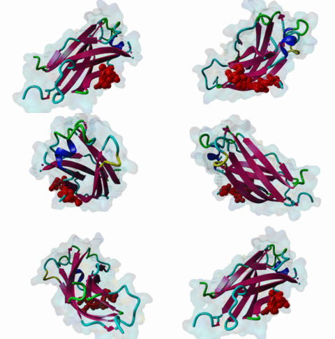
| Accession: | |
|---|---|
| Functional site class: | USP7 binding motif |
| Functional site description: | USP7, also known as HAUSP, is a deubiquitinating enzyme that cleaves ubiquitin moieties from its substrates. The USP7-mediated deubiquitination of p53, MDM2 and USP7 inhibition by the herpes viral proteins EBNA1 and ICP0 shows its importance in the regulation of cell survival pathways and in controlling key cellular processes important for viral infection. The N-terminal MATH domain of USP7 is responsible for substrate recognition and nuclear localization while the catalytic core domain is required for the deubiquitinating activity. The C-terminal Ubl domain is responsible for several USP7 substrate interactions, including ICP0, GMPS, DNMT1 and UHRF1 leading to substrate stabilisation, USP7 translocation or activation |
| ELMs with same func. site: | DOC_USP7_MATH_1 DOC_USP7_MATH_2 DOC_USP7_UBL2_3 |
| ELM Description: | Targeting motif found in a USP7 binding protein, docking to the MATH. The USP7 MATH domain is a TRAF-like domain but with different sequence specificity to the classical TRAF domain. The motif identified in EBNA1 recognises the same surface groove in USP7 as the p53 and MDM2 motifs, but requires a different description. The EBNA1 motif is high affinity but currently only this single example is known on which to base the motif. From the complex structure, core residues for the motif are P.E..S where the E substitutes for the P in the alternative motif (The E reaches to a complementarily-charged residue). It is not clear if there are cellular USP7 inhibitor (or substrate) proteins with the viral-like motif although this seems quite feasible. |
| Pattern: | P.E[^P].S[^P] |
| Pattern Probability: | 0.0007169 |
| Present in taxon: | Eukaryota |
| Interaction Domain: |
MATH (PF00917)
MATH domain
(Stochiometry: 1 : 1)
|
The DUB ubiquitin specific protease 7 (USP7) is a cysteine protease that was first identified as a binding partner for the Herpes simplex viral protein infected cell protein 0 (ICP0) which is why it is also called HAUSP (herpes simplex virus associated ubiquitin-specific protease). USP7 is responsible for the deubiquitination of many cellular proteins, thereby playing an important role in regulating biological processes including tumour suppression, immune responses, DNA repair and epigenetic control. (Nicholson,2011) Next to its catalytic domain, USP7 contains a N-terminal MATH domain and five C-terminal ubiquitin-like domains (UBLs) forming the HUBL domain (Faesen,2012). One of the most important USP7-interactions is the regulation of the tumour suppressor p53 through the stabilisation of MDM2 (HDM2), an E3 ligase which ubiquitinates p53 and thereby mediates its degradation (Nicholson,2011). MDM2 and p53 contain at least two binding sites for USP7, one on the N-terminal USP7 MATH domain, responsible for its nuclear localisation, and one on the C-terminal USP7 Ubl123 domain responsible for MDM2-catalysed p53 deubiquitination. Structural and mutational binding studies revealed that short peptides in disordered regions of both p53 and MDM2 interact with the same location on the USP7 MATH domain in a mutually exclusive manner (Ma,2010). The motifs responsible for N-terminal USP7 binding are based on a conserved P..S core, although the MDM2 variant binds USP7 with a much higher affinity possibly required for the role of USP7 in regulating p53 destruction by the MDM2 pathway (ELM: DOC_USP7_MATH_1, DOC_USP7_MATH_2) The motifs responsible for C-terminal USP7 interactions of MDM2 and p53 are still to be characterised. The MATH domain can also interact with the EBV protein EBNA1 to block USP7 activity and promote p53 elimination, as part of the mechanism to immortalise B-Cells (Saridakis,2005) The high affinity EBNA site approximates to a P.E..S motif. The Ser is equivalent in both motif classes and is suitable to be regulated by phosphorylation as a phospho Ser is structurally disallowed in the complex. However proline-directed kinases (MapKs, CDKs, Hipk2 etc.) could not phosphorylate these sites as Pro is structurally disallowed in the position following the Ser. The viral E3 ligase ICP0 binds to the C-terminal USP7 Ubl2 domain between amino acids 599-801 leading to a promiscuous activation of gene expression and the destruction of multiple cellular proteins, thereby overcoming the intrinsic cellular antiviral response (4WPH, 4WPI). The motif is characterised as a conserved K…K (DOC_USP7_Ubl2_3) core although the binding event might be regulated by cooperative binging of the N-terminal motif. This might explain how despite its low discrimination this binding event is able to maintain specificity. GMPS and UHRF1 also bind to the same USP7 binding pocket as ICP0, although this fact is still to be confirmed via crystal structures of these interactions. GMPS is involved in p53 regulation and the UHRF1-DNMT1-USP7 complex affects DNA methylation. (Pfoh,2015) DNMT1 was also shown to C-terminally bind USP7, using the same binding motif as ICP0 (4YOC, Cheng,2015). |
(click table headers for sorting; Notes column: =Number of Switches, =Number of Interactions)
| Acc., Gene-, Name | Start | End | Subsequence | Logic | #Ev. | Organism | Notes |
|---|---|---|---|---|---|---|---|
| P03211 EBNA1 EBNA1_EBVB9 |
442 | 448 | QGPADDPGEGPSTGPRGQGD | TP | 5 | Human herpesvirus 4 (strain B95-8) (Epstein-Barr virus (strain B95-8)) |
Please cite:
ELM-the Eukaryotic Linear Motif resource-2024 update.
(PMID:37962385)
ELM data can be downloaded & distributed for non-commercial use according to the ELM Software License Agreement
ELM data can be downloaded & distributed for non-commercial use according to the ELM Software License Agreement

